-
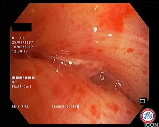
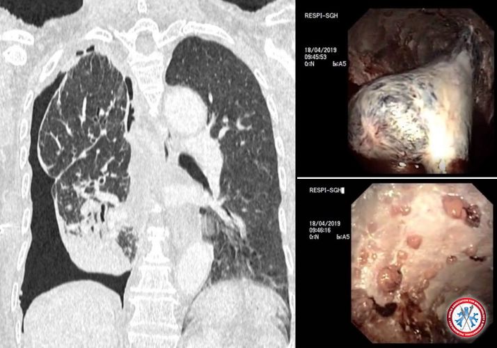
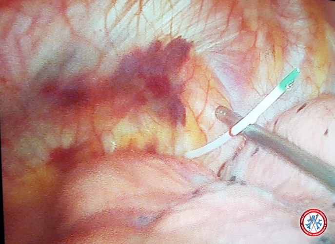
77-year-old female, non smoker, no known exposure who comes in with dyspnea, weight loss, left sided pleural effusion and pleural disease on chest CT. She underwent rigid pleuroscopy, fluid drainage, pleural biopsy and tunneled pleural catheter placement - Pathology: Stage IV Adenocarcinoma
Image courtesy of MOUNIR FERTIKH
5dd4324709d3d ADENOCA
-
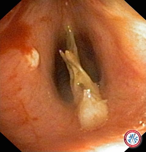
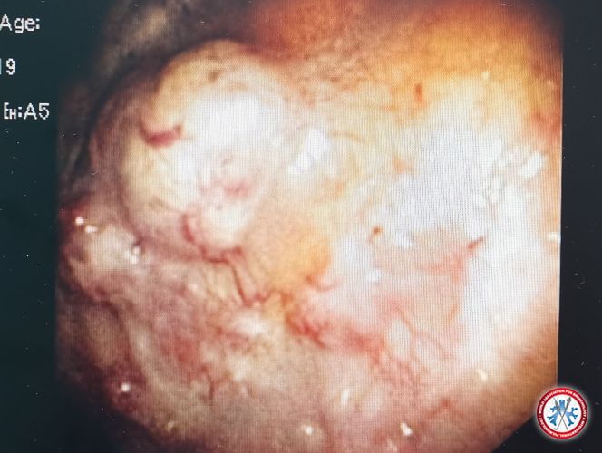
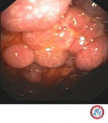
Malignant Trapped Lung - Metastatic Adenocarcinoma
Image courtesy of Kho Sze Shyang
5dd6207294516 Malignant trapped lung IMAGE 03
-
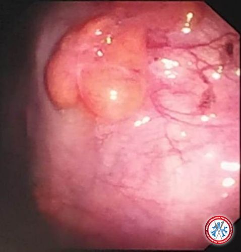
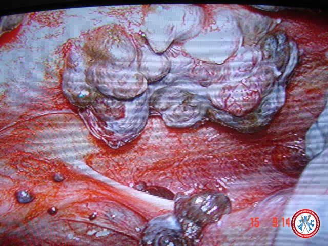
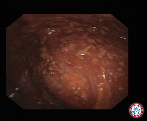
Multiple large nodules and superficial dilated vessels over parietal pleura
HPE: metastatic adenocarcinoma
Image courtesy of Lee Kai Quan
5ddfa58028ba6 IMG 20191124 080341 11694
-
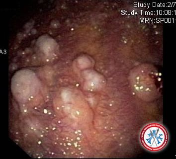
Pleural lipomatosis as a cause of pleural effusion --
Lipomas are benign tumors most commonly found in the subcutaneous tissue; however rarely, they may involve parietal pleura. Pleural lipomas are slow-growing tumors with no malignant potential but can diagnostic dilemmas to the clinicians. Pleural lipomas are benign soft-tissue neoplasms that originate from the submesothelial layers of parietal pleura and extend into the subpleural, pleural, or extrapleural space. They are soft, encapsulated fatty tumors with slow growth. The patient presented with pleural effusion of unknown etiology and semi rigid thoracoscopy revealed smooth, lobulated, multiple smooth yellowish nodules studded on the parietal pleura at multiple sites .Histopathology of biopsy sample was reported as adipose tissue comprising adipocytes and no evidence of atypia or malignancy, a finding which is characteristic of pleural lipomas.
Image courtesy of Saurabh Karmakar
5de38a58a66c8 Prebiopsy Copy
-
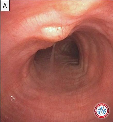
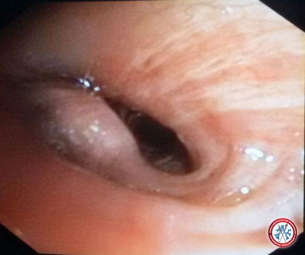
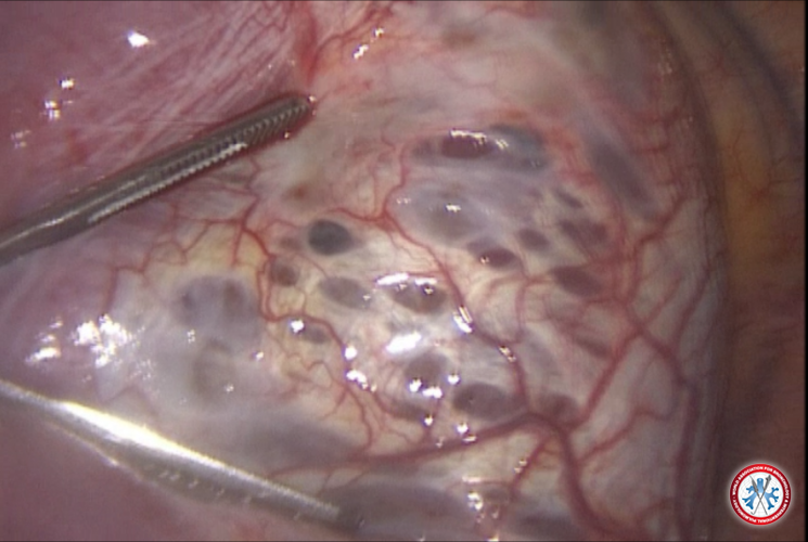
Right-sided diaphragmatic pores in a patient with catamenial pneumothorax.
Image courtesy of Liu Estradioto and Rodrigo Bettega de Araújo
5da689806fbf7 FIGURA 6
-
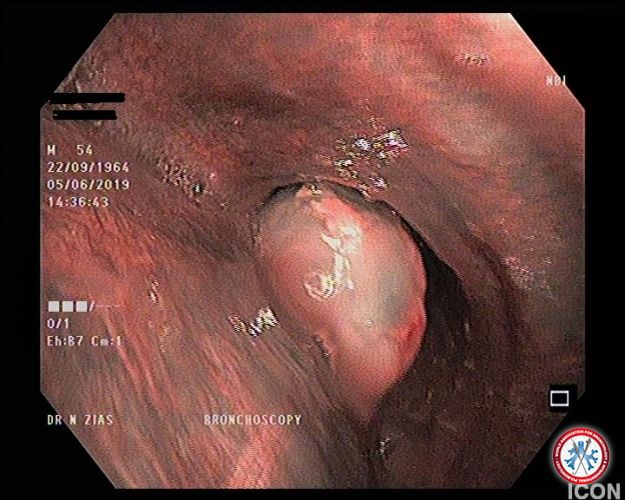
Thoracoscopic vision of central venous catheter wrongly positioned intrapleuraly.
Image courtesy of Cesar Ribeiro Zuccoli and Rodrigo Bettega de Araújo
5da689807078d FIGURA 9
-
Thoracoscopic image of parietal pleura showing potato like nodules- metastatic adenocarcinoma
Image courtesy of
Dr. Sharad Joshi
imagecontest wabip
-
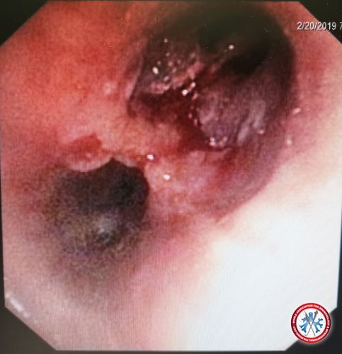
Case Metastatic Malignant Melanoma. Male Patient 45 years old , with history of malignant Melanoma of the skin , removed surgically and received chemotherapy.
2 years later , he had right sided pleural effusion , exaudate , on thoracoscopic examination we found multiple parietal pleural based dark black nodules of different sizes , biopsy revealed Malignant Metastatic Melanoma
Image courtesy of
Ayman A Hamid Farghaly, MD
imagecontest DSC01283
-
Multiple large whitish nodules over visceral and parietal pleura
HPE: metastatic adenocarcinoma
Image courtesy of
Lee Kai Quan, MD
imagecontest pleuro 2
-
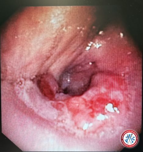
Hemorrhagic pleural effusion
Multiple whitish nodues over the parietal pleura
HPE: metastatic adenocarcinoma
Image courtesy of
Lee Kai Quan, MD
imagecontest pleuro 1
-
Thoracoscopy images of left pleural cavity with parietal pleura studded with large irregular nodules with yellowish tip - histopathology shown clear cell renal cell carcinoma - Fuhrman grade III.
Case description:
A 66 years old male patient with history of left nephrectomy 2 years back due to renal cell carcinoma, presented with left sided pleural effusion - and was found to have isolated left pleural metastasis of clear cell renal cell carcinoma.
Image courtesy of Dr. Jaykumar Mehta
20181027 191523
-
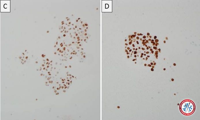
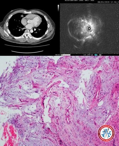
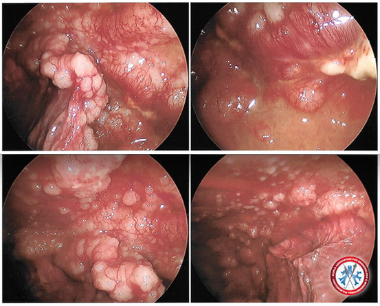
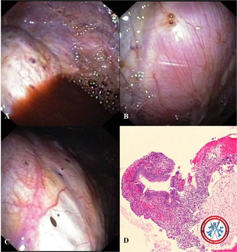
Pleuroscopic images demonstrate bloody pleural effusion and multiple small round red nodules on the diaphragm (A) and parietal pleura (B). Multiple small holes (*) are also discovered on the diaphragm (C). The histopathology of the nodules shows proliferative endometrial-typed glands surrounding by endometrial stroma which is covered with mesothelial cells (D). Thus, the diagnosis of pleural endometriosis is made by flex-rigid pleuroscopy.
Image courtesy of
Viboon Boonsarngsuk, MD & Tharintorn Chansoon, MD
imagecontest Fig 1
-
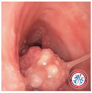
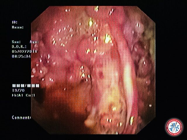
65yrs male patient with left sided encysted pleural effusion under gone thoracoscopy biopsy showing multiple grape like vascular nodules on parietal pleura. Biopsy revealed Squamous Cell carcinoma.
Image courtesy of
DR SOMNATH BHATTACHARYA
imagecontest DR SOMNATH THORACOSCOPY SQUAMOUS CA
-
Genitourinary (renal cell carcinoma)
Image courtesy of Fabien Maldonado, MD
5500cc5c0ed42 RCC pleura


