-
5dd47aca5f899 Slide 3 2
5dd47aca5f899 Slide 3 2
-
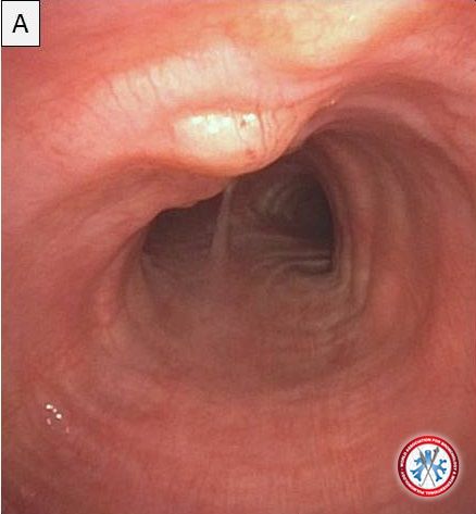
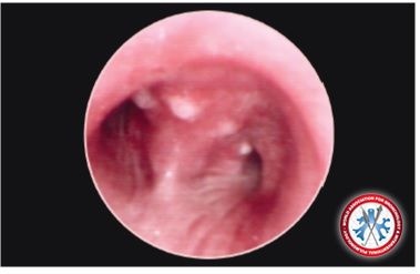
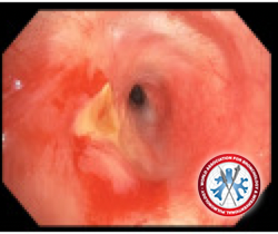
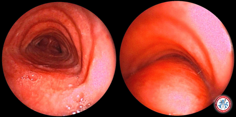
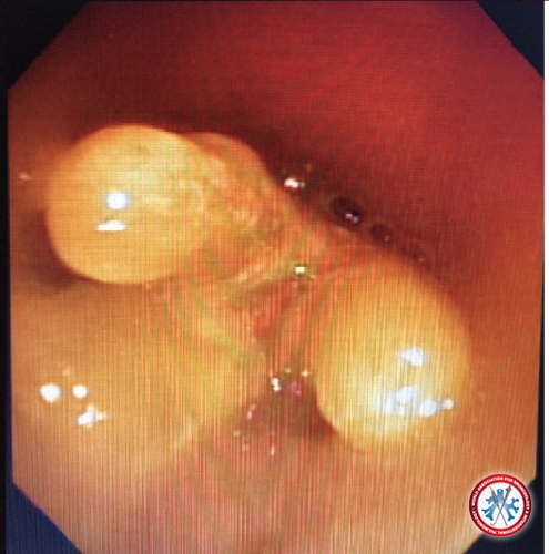
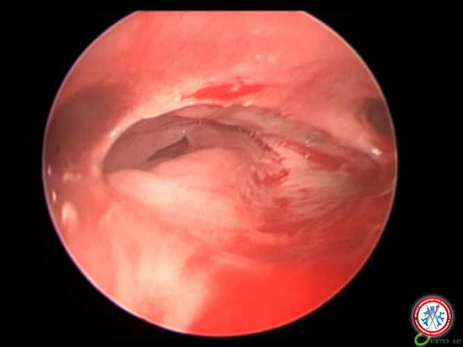
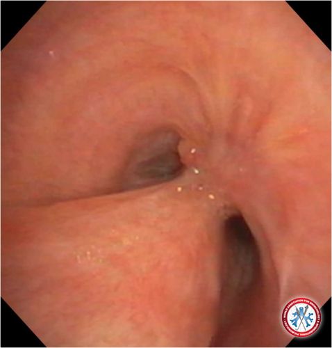
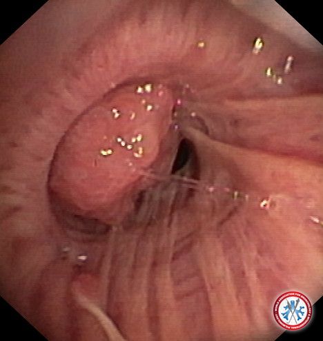
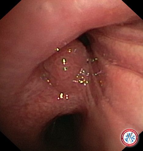
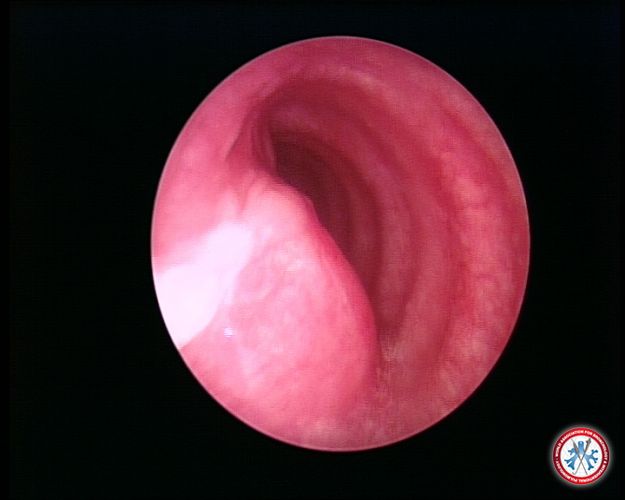
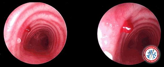
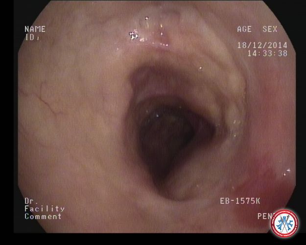
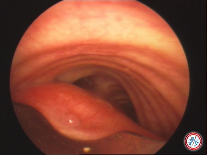
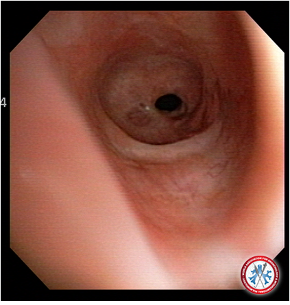
Figure A shows nodules anteriorly on the tracheal rings which may be mistaken for tracheobronchopathia osteochondroplastica (TPO) as it spares the posterior membrane.
Image courtesy of See Wei Low
5dd47aca5cae8 Slide1 1
5dd47aca5cae8 Slide1 1
-
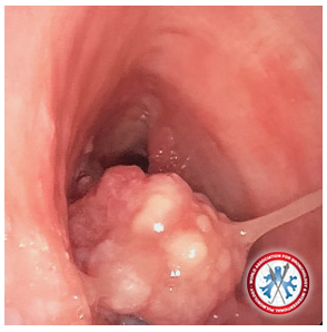
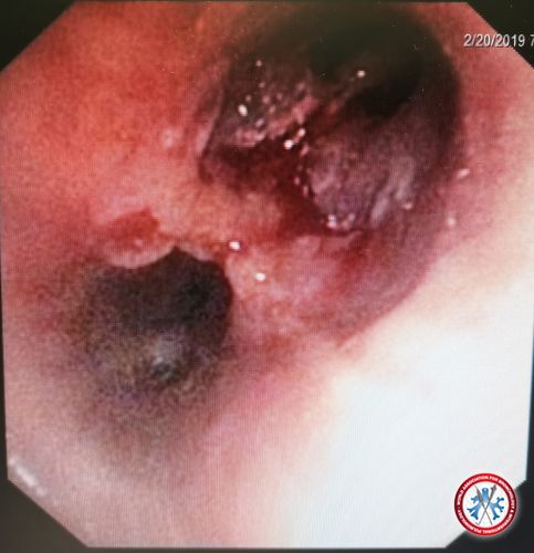
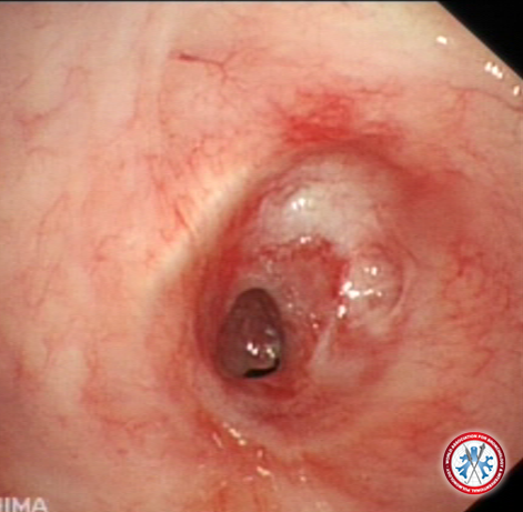
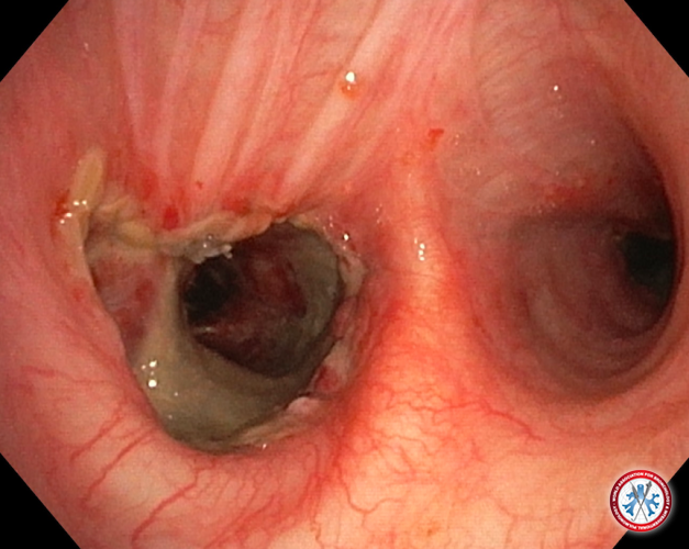
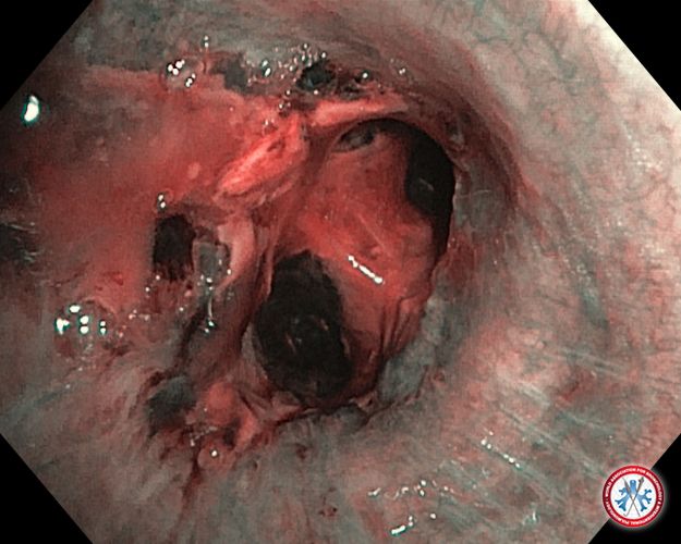
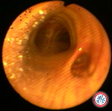
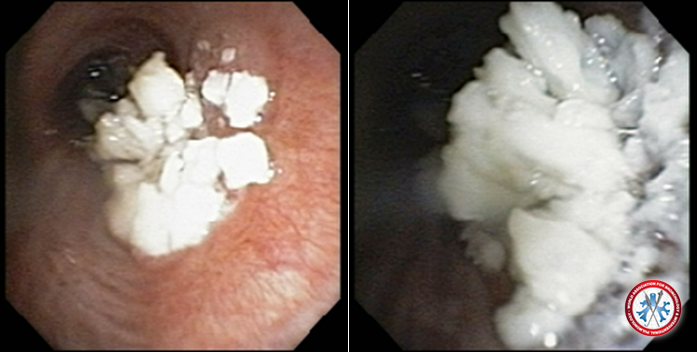
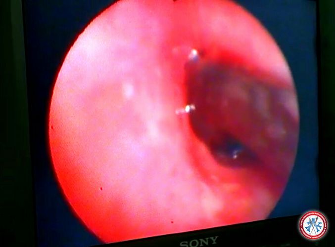
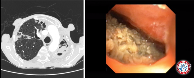
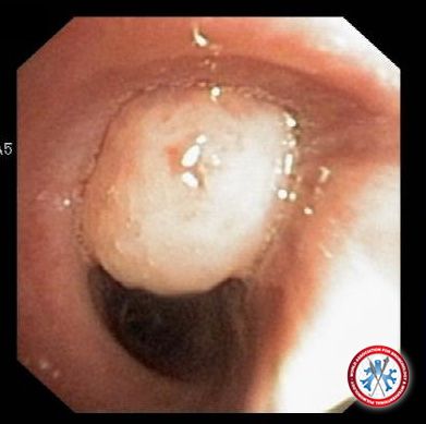
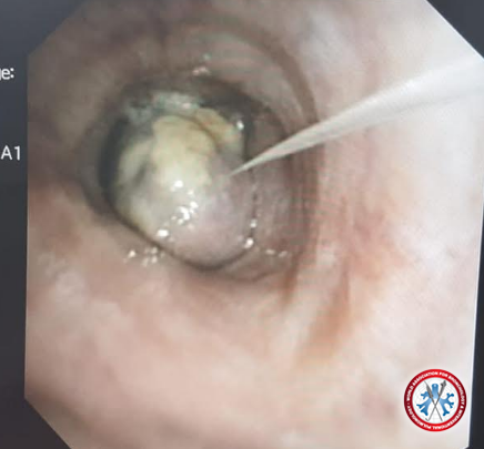
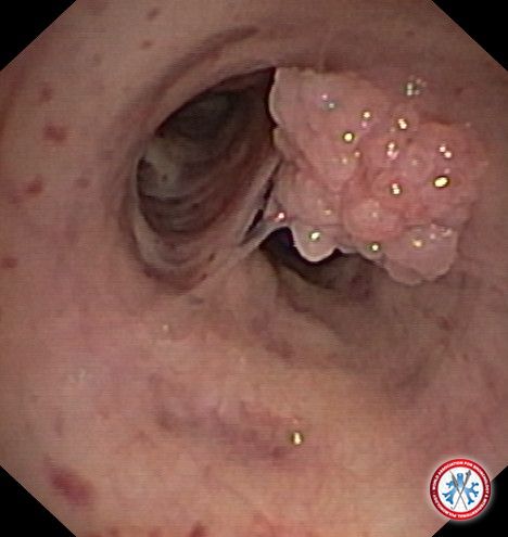
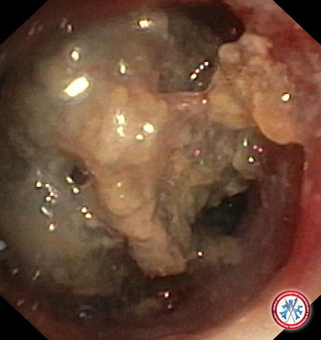
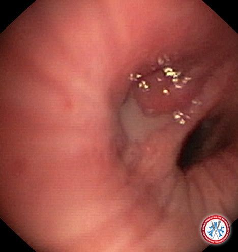
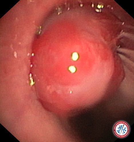
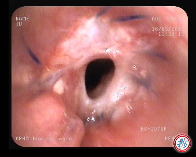
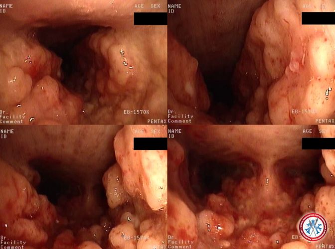
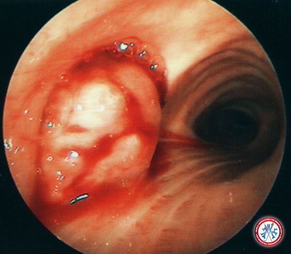
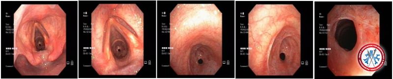
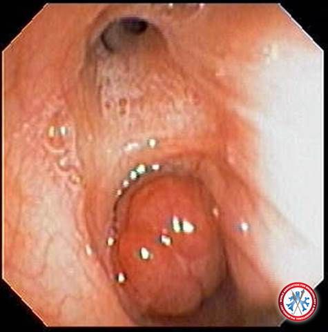
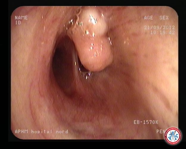
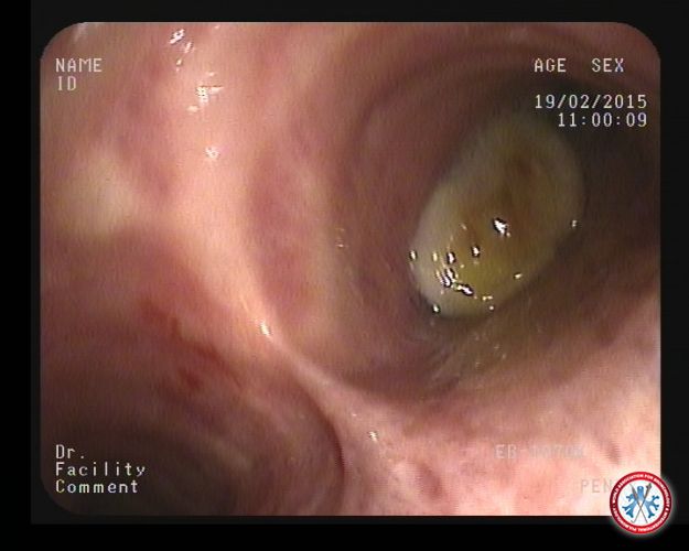
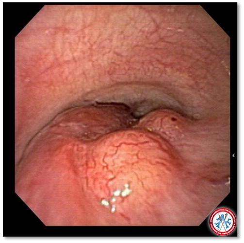
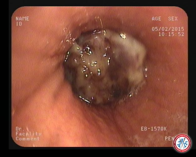
39 year-old-male, smoker who comes in with worsening dyspnea and mass noted on CT chest occluding the distal tracheal. Bronchoscopy with biopsy diagnosed papillomatosis.
Image courtesy of MOUNIR FERTIKH
5dd432470b364 RRP
5dd432470b364 RRP
-
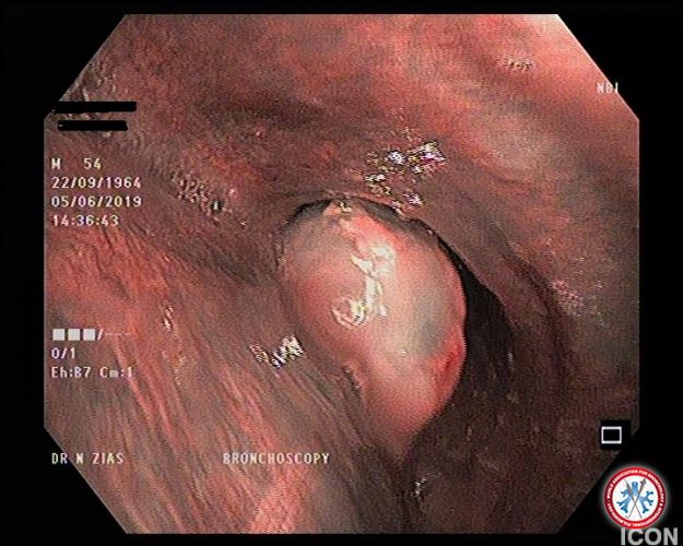
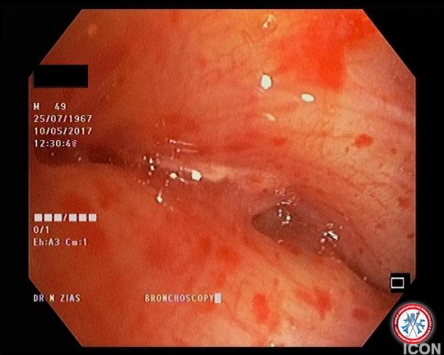
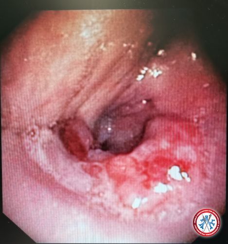
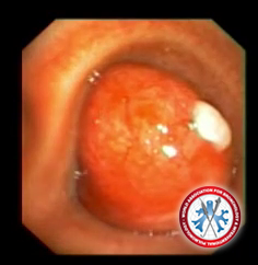
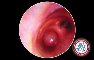
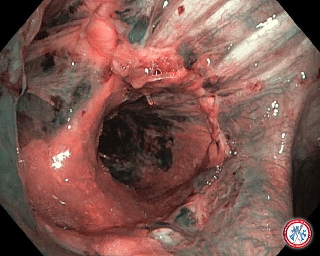
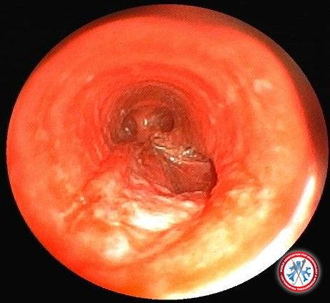
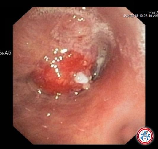
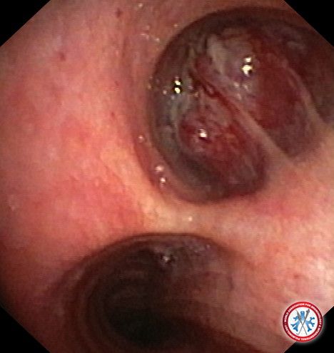
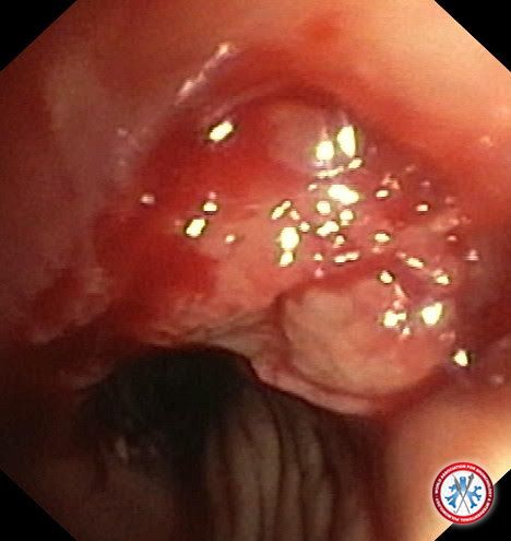
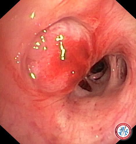
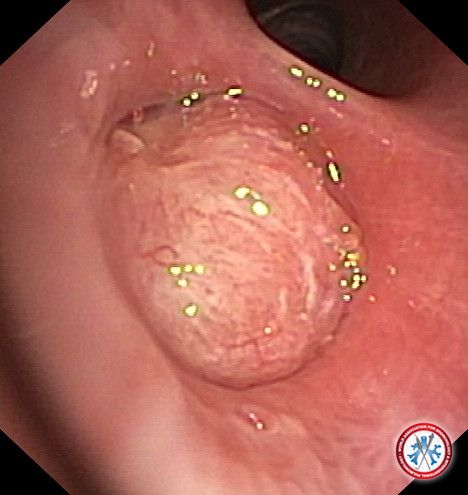
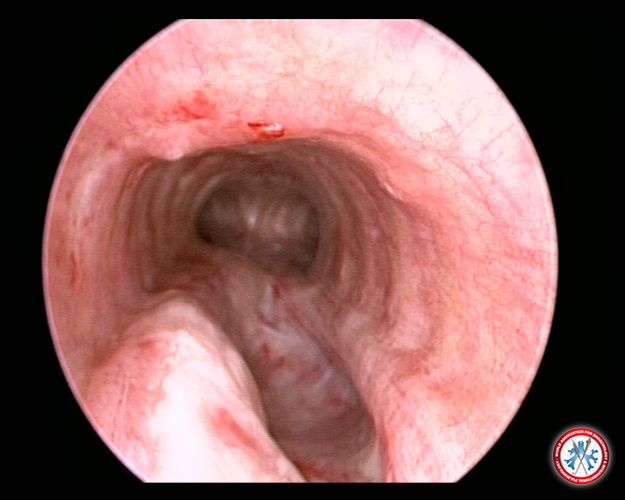
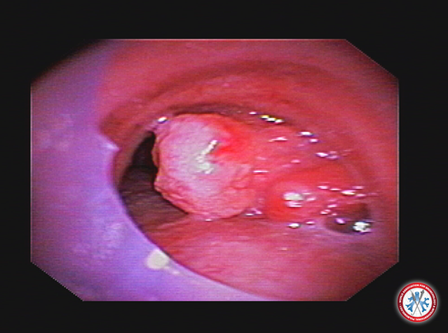
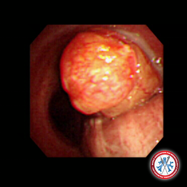
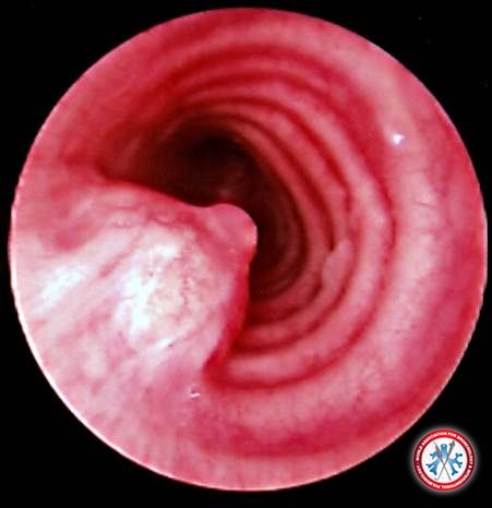
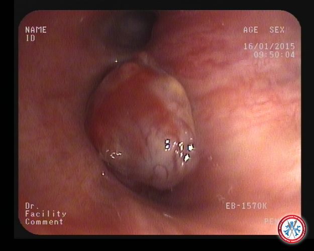
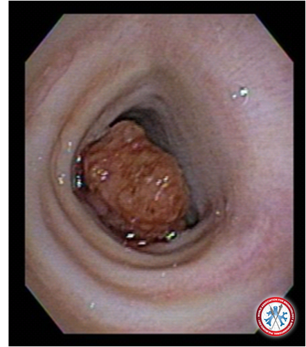
54 year old male patient presented with left main bronchus mass. Narrow-band bronchoscopy image. Biopsy revealed carcinoid tumor
Image courtesy of Zias Nikolaos
5dd3ca4279009 carcinoid narrow band
5dd3ca4279009 carcinoid narrow band
-
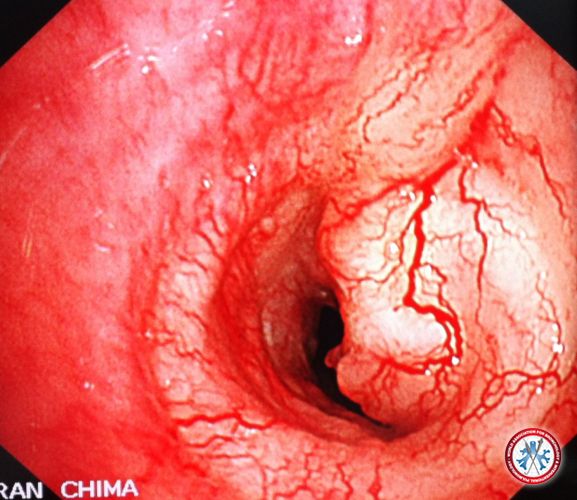
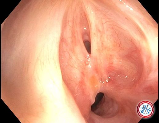
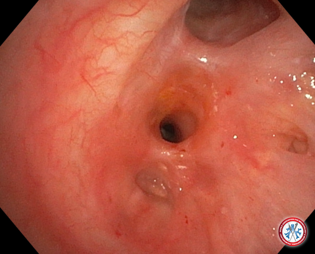
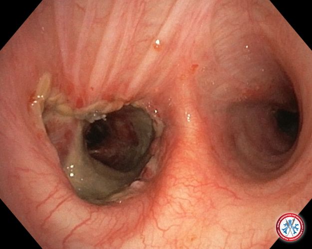
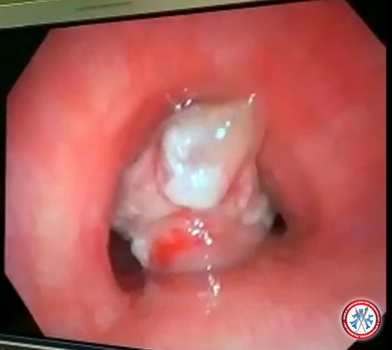
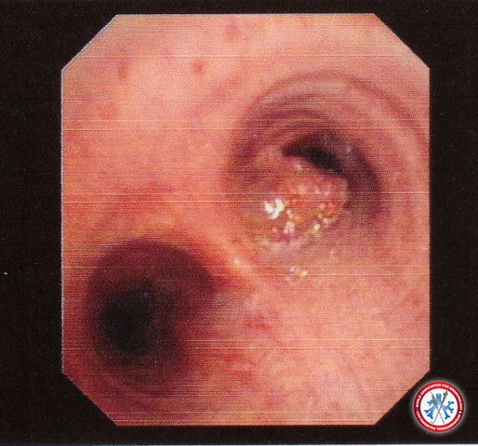
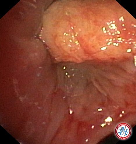
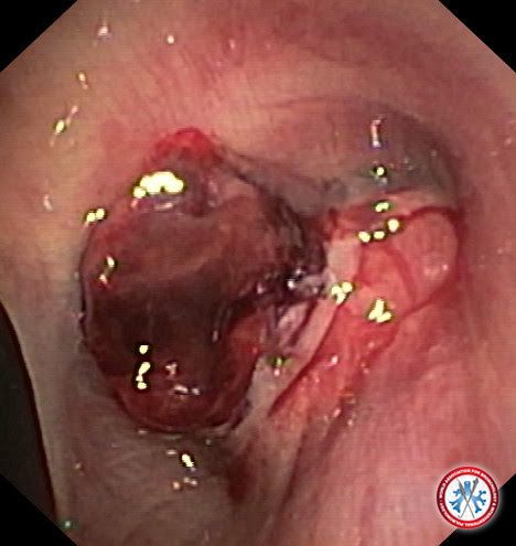
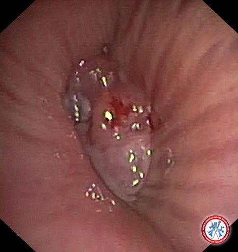
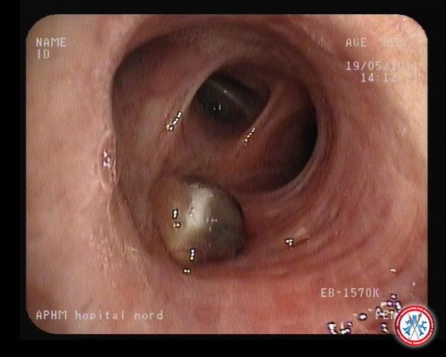
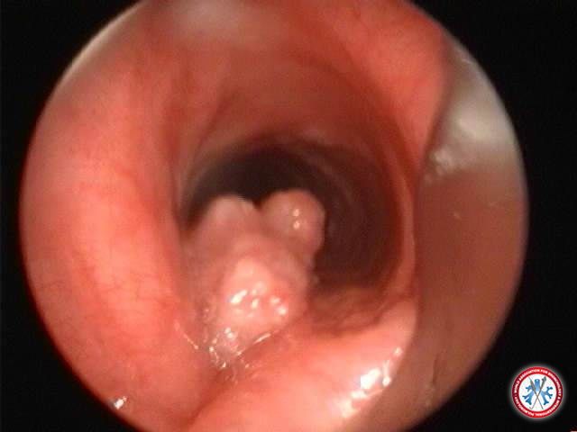
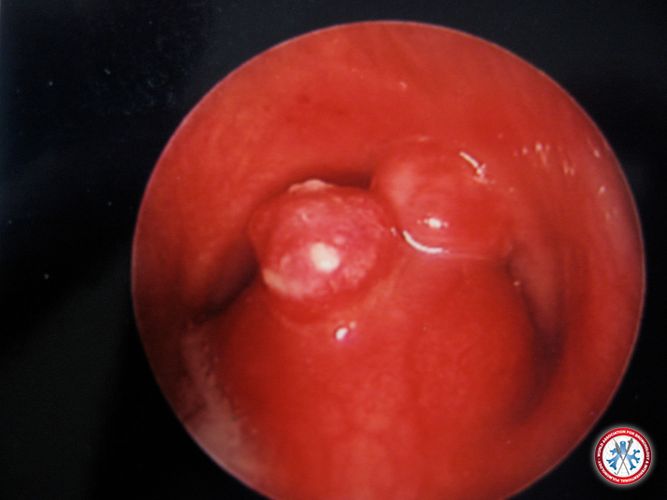
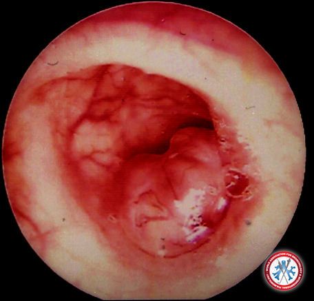
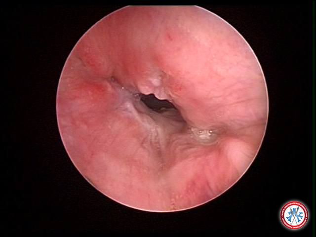
a 60-year old male patient with chronic cough, bronchoscopic image, biopsy revealed metastatic adamantinoma
Image courtesy of Apostolos Frimas
5dd3c66ae1b1b wabib image 1
5dd3c66ae1b1b wabib image 1
-
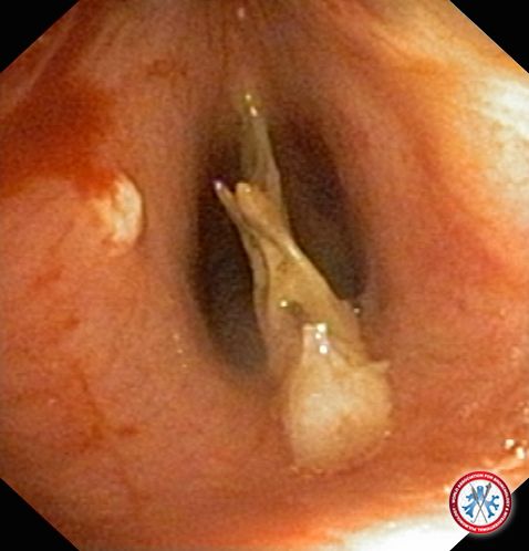
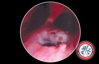
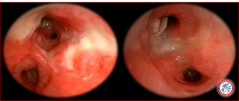
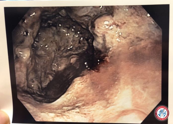
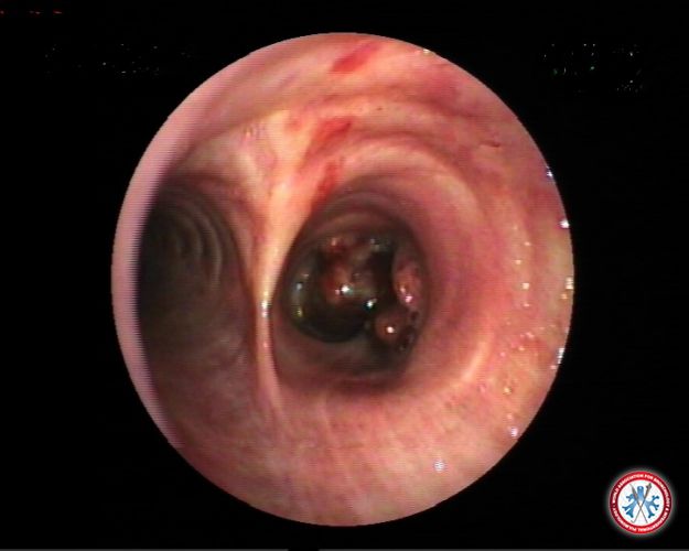
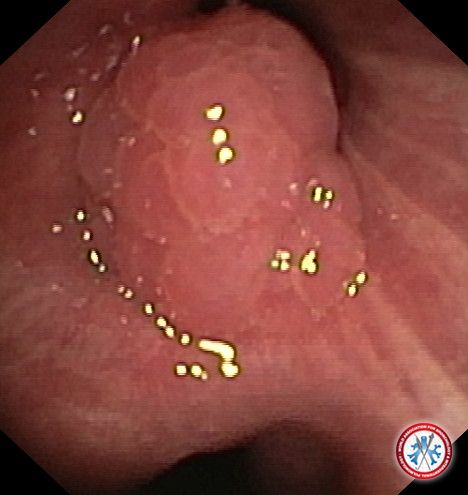
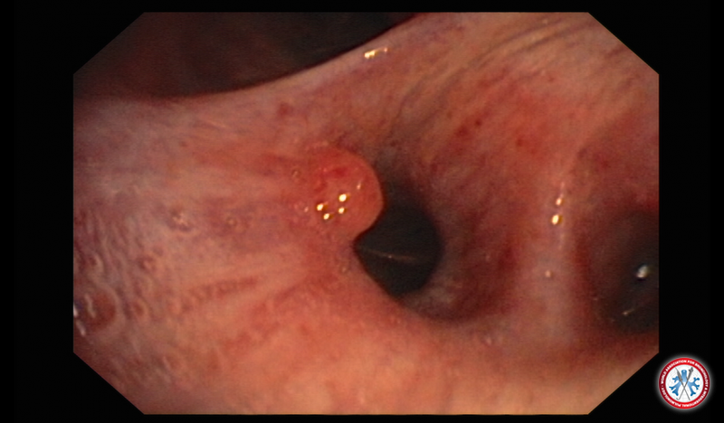
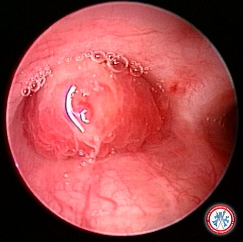
a case of a 35-year old male who presented with pain in the throat, inability to talk and wheezing while eating fish. The bronchoscopy revealed a fishbone placed perfectly between the vocal cords. The bone was snapped with forceps, then removed completely
Image courtesy of Apostolos Frimas
5dd3c66ae3f2d OLYM0007
5dd3c66ae3f2d OLYM0007
-
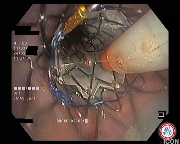
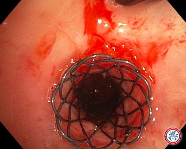
two endotracheal stents placed in a sleeve-like manner , required to provide adequate endotracheal pressure to open a left bronchus of a patient closed over 90% by mediastenum mass
Image courtesy of Apostolos Frimas
5dd3c66ae3b84 stent in stent
5dd3c66ae3b84 stent in stent
-
Endobronchial Melioidosis
Middle age gentleman from melioidosis endemic region presented with acute onset of fever with cough. CT thorax noted infiltrating mass at left hilar peri-aortic region. Bronchoscopy noted abnormal scaly mucosa at left main bronchus circumstantially on white light and narrow band imaging. Blood culture grew burkholderia pseudomallei and endobronchial biopsy consistent with inflammatory changes. Patient responded to prolonged antimicrobial and lesion resolved on surveillance imaging.
Image courtesy of Kho Sze Shyang
5dd6207292207 Endobronchial Melioidosis IMAGE 01
5dd6207292207 Endobronchial Melioidosis IMAGE 01
-
Pulmonary Carrtilaginous Hamartoma
Middle age gentleman presented with solitary pulmonary nodule in medial segment of right middle lobe during a routine chest radiograph. R-EBUS demonstrated an eccentric lesion with multiple hyperdense shadow within the target lesion. Transbronchial lung biopsy was consistent with cartilaginous pulmonary hamartoma.
Image courtesy of Kho Sze Shyang
5dd6207293feb Pulmonary Hamartoma IMAGE 02
5dd6207293feb Pulmonary Hamartoma IMAGE 02
-
unhealthy mucosa at secondary carina left lung, contact bleeding
HPE: squamous cell lung carcinoma
Image courtesy of Lee Kai Quan
5ddfa580243e0 Squamous cell carcinoma
5ddfa580243e0 Squamous cell carcinoma
-
Tumour infiltration over the right primary bronchus with friable mucosa and contact bleeding
HPE: small cell lung carcinoma
Image courtesy of Lee Kai Quan
5ddfa580273e1 IMG 20191120 093706 11704
5ddfa580273e1 IMG 20191120 093706 11704
-
Subglottic Tracheal stenosis in a patient of Granulomatosis with polyangiitis (GPA), earlier known as Wegener's granulomatosis
A non smoker patient aged 28 years old presented with cough off and on, dyspnea on exertion , hoarseness of voice and off and on hemoptysis since 3 months . Chest xray PA view and CT thorax showed multiple infiltrates in the bilateral upper lobes. Serum PR3 ANCA was elevated. A fibreoptic bronchoscopy revealed a tracheal stenosis in the subglottic area . Approximately 15% to 25% of all GPA patients experience subglottic stenosis.
Image courtesy of Saurabh Karmakar
5de38a58a3ba6 13
5de38a58a3ba6 13
-
Endobronchial lipoma as a cause of chronic cough -
A non smoker male patient presented with dry cough off and on, with no diurnal or seasonal variation , over 10 years. The cough had increased in intensity and frequency since 1 year and there was no associated dyspnea or other symptoms. Chest xray PA, spirometry and HRCT thorax was normal. A fibreoptic bronchoscopy revealed a smooth, lobulated, non-friable endobronchial mass on the opening of the right middle lobe bronchus without any features of atelectasis of the distal lobes. Biopsy confirmed it as lipoma. Endobronchial lipomas account for 0.1%–0.5% of lung tumors. Diagnosis is often delayed due to the indolent nature of this tumor. Common symptoms include a persistent cough (81% of cases), chest pain and dyspnea, recurrent fever, pneumonia, and wheezing (if it obstructs the lumen significantly).
Image courtesy of Saurabh Karmakar
5de38a58a6b30 Endobronchial Lipoma
5de38a58a6b30 Endobronchial Lipoma
-
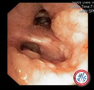
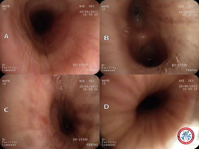
A severe distal left mainstem bronchial stenosis several months following a motor vehicle accident is shown in A1 where a pediatric bronchoscope could not be passed. After serial balloon dilation, the left upper and left lower lobes with cartilage fracture are shown (A2).
Image courtesy of See Wei Low
5dcce0f7374a3 A1A2
5dcce0f7374a3 A1A2
-
NSCCA lung on Histologypathology tumor invading upper 1/3rd of trachea with overlying vessels
Image courtesy of Dr. Muhammad Hussain
5dcaf0012ae3c IMG 1530
5dcaf0012ae3c IMG 1530
-
post intubation severe tracheal stenosis was manged with electrocautery knife dilatation. no recurrence till 3 years
Image courtesy of Dr. Muhammad Hussain
5dcaf0012e064 vlcsnap 2019 11 12 21h06m37s547
5dcaf0012e064 vlcsnap 2019 11 12 21h06m37s547
-
huge tumor occupying whole of upper trachea. was removed completely with cryo after resection electrocautery snare via Rigid bronchoscope. Vascular leiomyoma on H/P.
Image courtesy of Dr. Muhammad Hussain
5dcaf0012e81f vlcsnap 2019 11 12 22h15m20s400
5dcaf0012e81f vlcsnap 2019 11 12 22h15m20s400
-
Proximal airway of a 59 year-old female presenting with 3 months of progressive dyspnea following a non-tramautic intubation in the setting of surgical resection of the colon. In this picture, we are visualizing proximal subglottic stenosis that is actually tacking open the true vocal cords (also visualized).
Image courtesy of Daniel G. Dunlap
5dc44803633b9 SGS final
5dc44803633b9 SGS final
-
This patient, non-smoker, known case of diabetes mellitus not properly controlled for last 6 years, presented with dry cough, fever, breathlessness and loss of appetite for last 3 months. Computed tomography (CT) thorax showed multiple caseating mediastinal lymph nodes. Fiberoptic bronchoscopy showed fistula at lower end of trachea, surrounded by granulation tissue just before the main carina. Endobronchial biopsy from granulation tissue showed histopathology suggestive of tuberculosis. He was given antitubercular treatment (ATT) for 9 months along with antidiabetic treatment. Mediastinal lymph nodes disappeared and fistula also healed
Image courtesy of Dr Rajendra Prasad
5dc290c3b428b Tuberculous Lympho Tracheal Fistula in a 30 year old male
5dc290c3b428b Tuberculous Lympho Tracheal Fistula in a 30 year old male
-
This patient, bidi smoker (35/day for 45 years), a known case of COPD for last 7 years had presented with lung abscess like presentation with recurrent hemoptysis for last 6 months. Initially, he was misdiagnosed as pyogenic lung abscess and was prescribed antibiotic for 15 days with out any response. He went to another doctor, where he was again misdiagnosed as tuberculosis and was prescribed antitubercular treatment for 5 months with out any response, then he was referred to us. CT thorax showed mass in the right lung extending from the level of carina to level of diaphragm. Small, round, well-defined areas are seen anterior to the mass lesion and one of them was cavitating, suggestive of secondaries. Pleural effusion was also seen on right side. Fiberoptic bronchoscopy showed multiple nodule over main carina. Growth at the lateral wall of the right main bronchus obstructing right main bronchus partially. Growth was also extending to the medial wall of left main bronchus near main carina. Histopathology of endobronchial biopsy was consistent with squamous cell carcinoma
Image courtesy of Dr Rajendra Prasad
5dc290c3b681a SQUAMOUS CELL CARCINOMA IN A 65 YEAR OLD MALE
5dc290c3b681a SQUAMOUS CELL CARCINOMA IN A 65 YEAR OLD MALE
-
This patient, bidi smoker (5 bidi/day for last 6 years) presented with cough, fever, hemoptysis for last 1 year and was misdiagnosed as tuberculosis and was given antitubercular treatment for 1 year without any response. Fiberoptic bronchoscopy showed a polypoidal growth in left main bronchus. Growth was removed with the help of rigid bronchoscope. Biopsy of the growth revealed carcinoid tumor
Image courtesy of Dr Rajendra Prasad
5dc290c3b6500 CARCINOID TUMOR IN A 26 YEAR OLD MALE
5dc290c3b6500 CARCINOID TUMOR IN A 26 YEAR OLD MALE
-
anastomosis after lung transplantation. 3 week after LTx.
Image courtesy of Doctor Bekov Maksat
5db873e5d31c6 2019 10 29 20.06.28
5db873e5d31c6 2019 10 29 20.06.28
-
central air complication. 5 months after lung transplantation.
Image courtesy of Doctor Bekov Maksat
5db873e5d589f 2019 10 29 20.04.48
5db873e5d589f 2019 10 29 20.04.48
-
after stent installation. 6 months after transplantation.
Image courtesy of Doctor Bekov Maksat
5db873e5d6d1a 2019 10 29 20.05.33
5db873e5d6d1a 2019 10 29 20.05.33
-
right bronchial anastomosis in mode NBI.
Image courtesy of Doctor Bekov Maksat
5db8752627036 800H0011
5db8752627036 800H0011
-
lung transplant.
Image courtesy of Doctor Bekov Maksat
5db8752627f5a 800H0009
5db8752627f5a 800H0009
-
left bronchial anastomosis in mode NBI.
Image courtesy of Doctor Bekov Maksat
5db8752627840 800H0012
5db8752627840 800H0012
-
Left mainstem bronchial stenosis due to tuberculous with endobronchial component.
Image courtesy of Kim Styrvoky MD
5dae268256825 Screen Shot 2019 10 21 at 4.27.09 PM
5dae268256825 Screen Shot 2019 10 21 at 4.27.09 PM
-
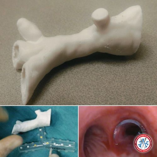
Airway stenting (Oki stent) guided by a 3D printed model of corrected airways (top). Modification of the stent with precise cutting of its different arms (right main bronchus, upper right lobe, bronchus intermedius) according to data from the 3D model (bottom left). Bronchoscopic views of the main right bronchus before and after stenting (bottom right)
Image courtesy of GUIBERT Nicolas
5dab8c06d8816 IMG 20191020 001058 357
5dab8c06d8816 IMG 20191020 001058 357
-
Saber-sheath trachea in a patient with COPD.
Image courtesy of Liu Estradioto, MD & Caroline Blum, MD.
5da5b73d8af4c saber
5da5b73d8af4c saber
-
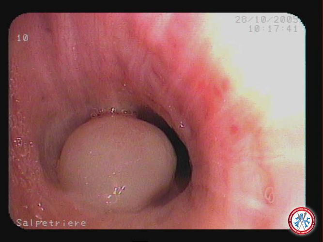
Accessory cardiac bronchus accidentaly discovered in a patient during a routine bronchoscopic for tracheostomy care.
Image courtesy of Rodrigo Bettega de Araújo
5da64b2f3ffc2 BRONQUIO CARDIACO ACESSORIO
5da64b2f3ffc2 BRONQUIO CARDIACO ACESSORIO
-
Seven centimeter long posterior tracheal wall rupture after emergency oral intubation.
Image courtesy of Rodrigo Bettega de Araújo
5da64b2f43519 LACERA O PAREDE POSTERIOR DA TRAQU IA
5da64b2f43519 LACERA O PAREDE POSTERIOR DA TRAQU IA
-
Tracheal bronchus arising close de the main carina on the right side of the tracheal wall.
Image courtesy of Liu Estradioto and Rodrigo Bettega de Araújo
5da688cf1f39f FIGURA 2
5da688cf1f39f FIGURA 2
-
Diverticulum on the lower trachea. Some authors believe that a former tracheal bronchus has either resorbed or not developed, leaving only a blind pouch remnant.
Image courtesy of Liu Estradioto and Rodrigo Bettega de Araújo
5da688cf2162e FIGURA 3
5da688cf2162e FIGURA 3
-
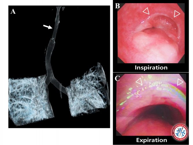
Expiratory Central Airway Dinamic Colapse in a patient with mild dyspnea, expiratory wheezing and intermitent stridor.
Image courtesy of Liu Estradioto and Rodrigo Bettega de Araújo
5da688cf21d64 FIGURA5
5da688cf21d64 FIGURA5
-
Seven centimeter long posterior tracheal wall rupture after emergency oral intubation.
Image courtesy of Liu Estradioto and Rodrigo Bettega de Araújo
5da68980714ec FIGURA 11
5da68980714ec FIGURA 11
-
White plaques and exofitic lesions on the apico-posterior bronchus of the left upper lobe in a patient with Mycobacterium tuberculosis and Candida sp coinfection.
Image courtesy of Liu Estradioto and Rodrigo Bettega de Araújo
5da68afc711eb FIGURA 14
5da68afc711eb FIGURA 14
-
A tooth founded inside the trachea above the tracheostomy tube during a routine bronchoscopy for tracheal canula change.
Image courtesy of Liu Estradioto and Rodrigo Bettega de Araújo
5da68afc6fccf FIGURA12
5da68afc6fccf FIGURA12
-
Squamous cell carcinoma of the left main bronchus. Note the hyperkeratinization.
Image courtesy of Liu Estradioto, MD & Rodrigo B. Araújo, MD.
5da0cc01e3a88 cec1
5da0cc01e3a88 cec1
-
A Case of Total Resection of Typical Endobronchial Carcinoid Tumor with Rigid Bronchoscopy
A Mimic of Asthma
We report a 25-year-old woman with persistent dyspnea and wheezes that had been unsuccessfully treated with inhaled beta 2-agonists and steroids for about one year. Spirometry demonstrated a restrictive pattern. Chest CT demonstrated polypoidal lesion in left main bronchus. The lesion was excised via rigid bronchoscopy. Pathology showed a picture of typical bronchial carcinoid. In this patient, due to the lack of awareness, diagnosis of carcinoid was delayed for one year.
Image courtesy of : Ayman Rizk
5d9d9955eb693 1 1
5d9d9955eb693 1 1
-
A Case of Total Resection of Typical Endobronchial Carcinoid Tumor with Rigid Bronchoscopy
A Mimic of Asthma
We report a 25-year-old woman with persistent dyspnea and wheezes that had been unsuccessfully treated with inhaled beta 2-agonists and steroids for about one year. Spirometry demonstrated a restrictive pattern. Chest CT demonstrated polypoidal lesion in left main bronchus. The lesion was excised via rigid bronchoscopy. Pathology showed a picture of typical bronchial carcinoid. In this patient, due to the lack of awareness, diagnosis of carcinoid was delayed for one year.
Image courtesy of Ayman Rizk
5d9d9955ed2a9 3 1
5d9d9955ed2a9 3 1
-
Preganant Lady with pulmonary tuberculosis intubated for respiratory distress. Chest xray revealed total left lung collapse. Bedside bronchoscope showed large blood clot occluding Left Main Bronchus. Removed via cryoprobe. Lung expanded and patient was extubated subsequently
Image courtesy of Arvindran Alaga
5d9d55e76ffa2 20191009 111633
5d9d55e76ffa2 20191009 111633
-
Patient presented with stridor. Noted to have tracheal mass. Debulking done via Snare and hemostasis secured via laser. Histopathological examination revealed metastatic renal cell carcinoma
Image courtesy of Arvindran Alaga
5d9d55e76f202 20191009 112024
5d9d55e76f202 20191009 112024
-
RUL cavity because of MSSA infection in lung cancer patient
Image courtesy of Dr. Khalid Al Efraij
5d9d3764d4cf4 563667BB B791 444F A8D5 C2318C6CFF61
5d9d3764d4cf4 563667BB B791 444F A8D5 C2318C6CFF61
-
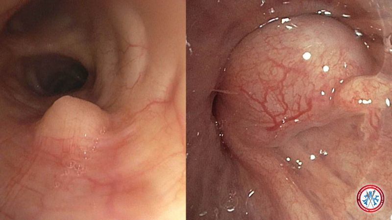
Comparing benign airway tumors - chondroma of the left mainstem bronchus (left image) and endobronchial hamartoma of the right upper lobe (right image)
Image courtesy of Dhaval Thakkar, MD, Carla Lamb, MD & Sara Shadchehr, MD
3
3
-
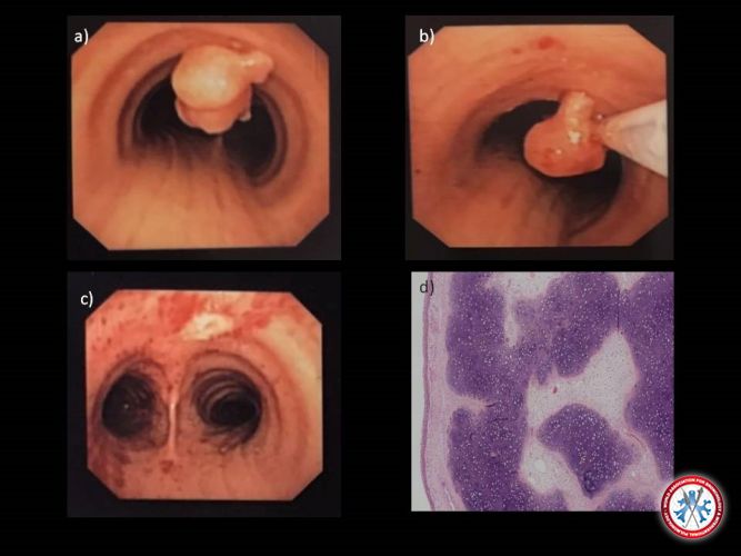
Endotracheal chondroma. a) A large polypoid mass at the anterior wall of the lower third of the trachea, b-c) successfully excised by snare electrocautery during flexible bronchoscopy. d) Lesion composed of hyaline cartilage and loose myxoid stroma under the normal bronchial mucosa; low cellularity and no atypia (Hematoxylin and eosin, X40)
Image courtesy of Ilias Porfyridis, M.D.
2
2
-
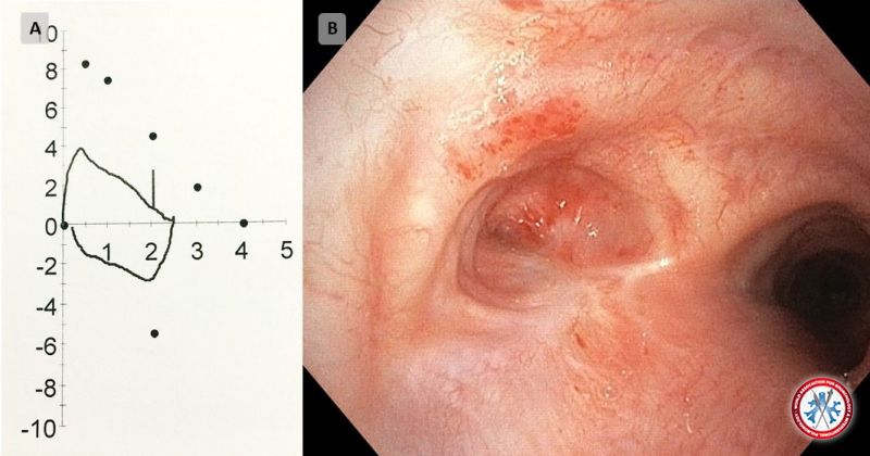
Refractory hydrothorax appears when there is no response to sait restriction, diuretics and paracentesis and its management is not well established. Videothoracoscopy is a promising therapy that permits the detection and closure of diaphragmatic defects, and when used with talc pleurodesis resulted in long-lasting control.
Image courtesy of Salvato Feijó, M.D.
1
1
-
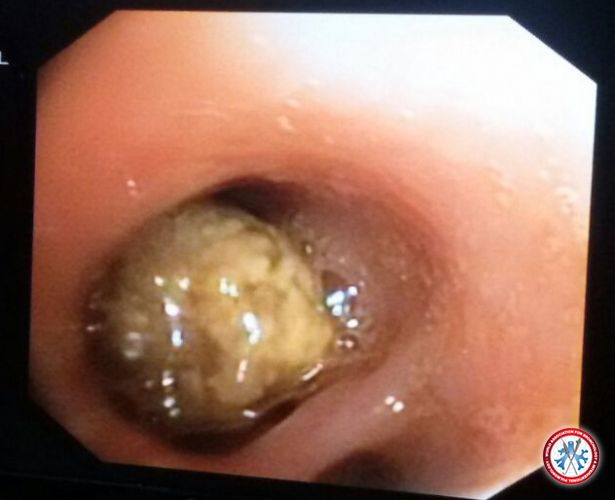
24-year-old female presented with complaints of massive hemoptysis since 1 year off and on. She was a life-long nonsmoker and there was no history of other illnesses. Sputum for acid-fast bacilli was negative. Sputum culture for bacteria and fungus was negative.
The patient's chest x-ray and HRCT thorax were normal. Flexible fibreoptic bronchoscopy was performed to evaluation of the airways. The bronchoscopy revealed an irregularly shaped yellowish mass of approximately 2-cm size partially obstructing the right middle lobe bronchus . The mass was lodged firmly to the mucosa and couldn't be dislodged. A few biopsies were taken and the histopathology revealed it to be aspergilloma.
Image courtesy of
Dr. Saurabh Karmakar
imagecontest Fungal Ball Copy
imagecontest Fungal Ball Copy
-
Adenoid Cystic Carcinoma endo-tracheal bud.
The endoscopy was done on a 25-year-old woman with a seven months’ history of asthma but whose radiological and endoscopic exploration revealed an intra tracheal bud of 2.5 cm in diameter sitting at 4 cm below the epiglottis. This formation is richly vascularized with easy bleeding. After histological confirmation the tumor was resected surgically
Image courtesy of
BENAZZOUZ REDOUENE SID AHMED, MD
imagecontest Cylindrome Red Benz
imagecontest Cylindrome Red Benz
-
A Migrating Foreign Body.
Patient presented with right pyopneumothorax with hyperdense lesion seen within the distal bronchus intermedius. Bronchoscopy noted granulation tissue at distal bronchus intermedius (Panel B) with no intraluminal lesion. Further examination noted the foreign body was migrated to the left lower lobe bronchus (Panel C) which was removed successfully via rigid bronchoscope. Patient revealed a prior history of choking on chicken bone.
Image courtesy of
Kho Sze Shyang, MD
imagecontest IMAGE 03
imagecontest IMAGE 03
-
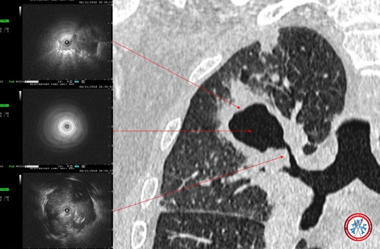
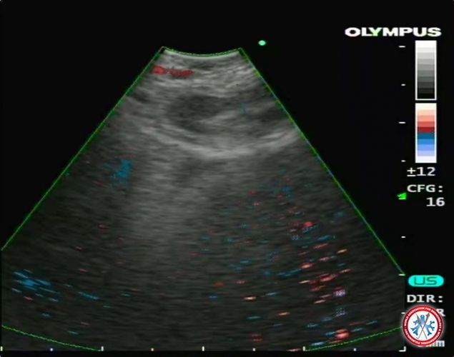
Anatomy of A Malignant Pulmonary Cavity.
Radial endobronchial ultraosund (R-EBUS) image of thick walled pulmonary cavity at various contact point. Biopsy proven adenosquamous carcinoma of the lung.
Image courtesy of
Kho Sze Shyang, MD
imagecontest IMAGE 01
imagecontest IMAGE 01
-
Tracheoesophageal fistula at the main carina
Image courtesy of
Evangelos Balis, MD
2018 12 06 19h29m48s112
2018 12 06 19h29m48s112
-
Huge pulmonary Aspergilloma: a crescent of air above a truffle-shaped mass that appeared mobile inside a neoformed cavity from a destroyed left upper lobe.
Image courtesy of
Rachid TAZI-MEZALEK, MD & Felipe ANDREO GARCIA, MD
imagecontest Aspergiloma Dr Tazi
imagecontest Aspergiloma Dr Tazi
-
A mulberry-like mass at the proximal part of right principal bronchus, partially occluding the lumen.
Image courtesy of
Nirmal Kanti Sarkar, MD
imagecontest Bronchial rhinosporidiosis
imagecontest Bronchial rhinosporidiosis
-
33 y male, Non-Hodgkin lymphoma
Image courtesy of
Ahmet ILGAZLI, MD
imagecontest Non Hodgkin lymphoma
imagecontest Non Hodgkin lymphoma
-
Biphasic Flow Volume Loop in Post Tuberculous Central Airway Obstruction
A young patient was referred to us for complete left lung collapse after completion of anti tuberculous therapy. He demonstrated microbiological and clinical improvement despite the worsening radiographic changes. Spirometry show biphasic flow volume loop and bronchoscopy noted an obliterated left main bronchus ostium with whitish fibrotic plaque consistent with post tuberculosis changes. Subsequent computed tomography of the chest shown total left lung destruction which was not feasible for airway intervention.
Image courtesy of
Kho Sze Shyang, MD
imagecontest IMAGE 04
imagecontest IMAGE 04
-
Cupid's arrow-EBUS-TBNA for 4R.
The patient was diagnosised as NSCLC with biopsies from TBLB and TBNA. The picture shows the process of EBUS-TBNA on 4R lymph node. On the right upper site of the picture, we can see the needle inserted into the heart-shaped lympn node, so we call it Cupid's arrow.
Image courtesy of Xiaolian Song, MD
Cupid s arrow EBUS TBNA for 4R
Cupid s arrow EBUS TBNA for 4R
-
Sarcoidosis- confocal image of the right upper bronchial mucosa. The patient was diagnosised as Sarcoidosis with biospies from the airway wall. The picture is one of the confocal images of the right upper bronchial mucosa, which diplays highlight spots and disorderly arranged fibers of the airway wall under the confocal probe.
Image courtesy of Xiaolian Song, MD
Sarcoidosis confocal image of the right upper bronchial mucosa
Sarcoidosis confocal image of the right upper bronchial mucosa
-
Bronchoscopic image showing High attenuation mucous in left main bronchus in a case of ABPA
Image courtesy of
Dr. Abhisheka Kumar.
imagecontest IMG 20181027 142101 01 02
imagecontest IMG 20181027 142101 01 02
-
A 26-year-old male presented with a two years history of intermittent chronic cough, sore throat, weight loss, dysphonia, he had low grade of fever associated with gradual deterioration in health.
Bronchoscopy was done: smear positive and biopsy the histopathological examination revealed Langhan’s type giant cells surrounded by lymphocytes and fibroblasts with a few areas of caseous necrosis suggestive of tuberculosis.
Image courtesy of
Danny Alvarado Maldonado, MD
imagecontest Laryngeal TB
imagecontest Laryngeal TB
-
Tracheal stenosis post radiation for squamous cell ca forming a pseudo carina.
Image courtesy of
YIPING FU, MD
imagecontest leddy pseudocarina
imagecontest leddy pseudocarina
-
Extensive nodular lesions on entrance of right main bronhus, extending to right intermediate bronchus, minima contact bleeding on biopsy
HPE: Adenicarcinoma of lungs
Image courtesy of
Lee Kai Quan, MD
imagecontest broncho 2
imagecontest broncho 2
-
Right upper lobe Total obstruction by necrotic tumour
HPE: Adenocarcinoma of lungs
Image courtesy of
Lee Kai Quan, MD
imagecontest broncho 33
imagecontest broncho 33
-
Left main bronchus, less than 2 cm from carina, dilated spuerfical vessels with contact bleeding on biopsy
HPE: Squamous cell carcinoma
Image courtesy of Lee Kai Quan, MD
imagecontest bronchi 19
imagecontest bronchi 19
-
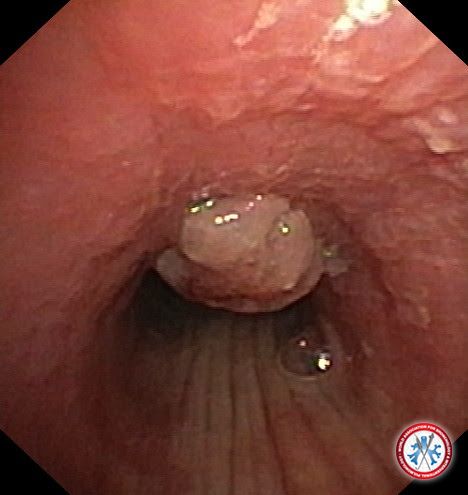
Carcinoid tumour obstructiong left main bronchus, 3cm distal to carina
Image courtesy of
Anita Graham, MD
imagecontest carcinoid tumour
imagecontest carcinoid tumour
-
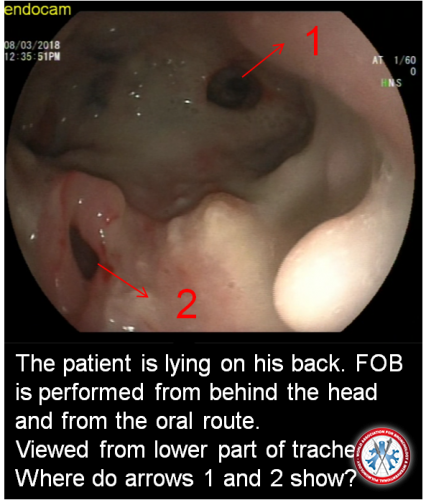
The patient is lying on his back. FOB is performed from behind the head and from the oral route.
Viewed from lower part of trachea. Arrow 1 shows intermedius bronchus and arrow 2 shows oesophagus.
Huge mediastinal/lung tumor had destructed lower part of trachea and mediastinal structure.
Image courtesy of
Onur Erer, MD
imagecontest Resim5
imagecontest Resim5
-
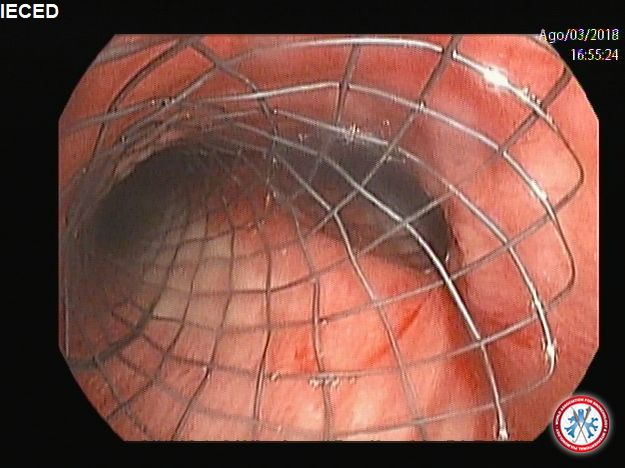
67 y/o Female heavy smoker with a large mass in lower trachea that cause extrinsic compression at the level of main carina. Two no cover SEMS stents they were placed with great clinical resolution.
Image courtesy of
Gonzalo Ugarte Fornell, M.D.
imagecontest STENT X 2
imagecontest STENT X 2
-
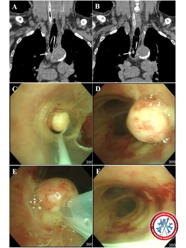
An 86 year-old woman with a chronic obstructive pulmonary disease was intubated and
admitted to intensive care unit for acute respiratory failure due to acute exacerbation. When
her respiratory distress was improved on the third ventilator day, ventilator weaning was
attempted. However, a spontaneous breathing trial with a T-piece was failed in a short period
of time. Though objective findings were compatible with weaning criteria, she still reported
dyspnea under weaning trial. Further evaluation by chest computed tomography revealed a
mass located in the distal end of endotracheal tube (Panel A and B, arrow). She underwent
flexible bronchoscopy, which revealed endotracheal polyp occlude distal opening of
endotracheal tube (Panel C). The polyp was resected endoscopically with using snare (Panel
D, E and F). Pathology revealed fibro-epithelial polyp, an extremely rare type of benign tumor
in trachea. After resection of tracheal polyp, she was successfully extubated without respiratory
distress
Image courtesy of
Yousang Ko, MD
imagecontest WABIP unusual weaning failure
imagecontest WABIP unusual weaning failure
-
An 81-year-old woman had undergone salpingo-oophorectomy under general anesthesia. Five days after operation, she started complaining of dyspnea, but relived her symptom by coughing. Chest computed tomography with coronal reconstruction showed segmental luminal narrowing in the proximal trachea just below the vocal cords. (Figure 1A, arrow). She underwent flexible bronchoscopy, which revealed fibrinous tracheal pseudo-membrane (FTP) complicated after intubation. It dynamically obstructed her trachea by flapping, closed in inspiration and opened in expiration, so her symptom was relieved by coughing (Figure 1B, C, arrowhead). Unfortunately, she was worsened respiratory distress and intubated during bronchoscopy. After intubation with 6.0-mm tracheal tube, we inserted the flexible bronchoscope through remnant space of both vocal cords and separated that from cartilaginous portion of trachea using by biopsy forceps. Finally, her dyspnea resolved immediately after procedure.
Image courtesy of
Yousang Ko, MD
imagecontest WABIP tracheal pesudomemb
imagecontest WABIP tracheal pesudomemb
-
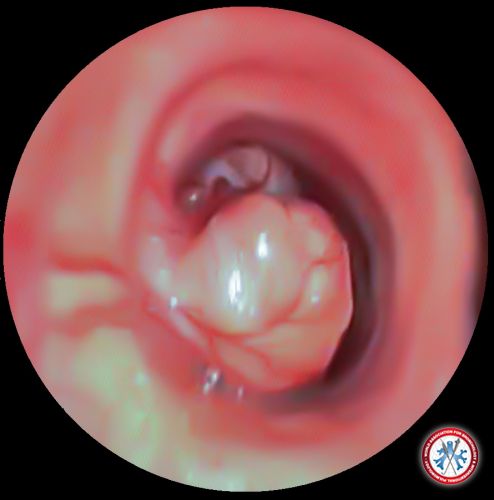
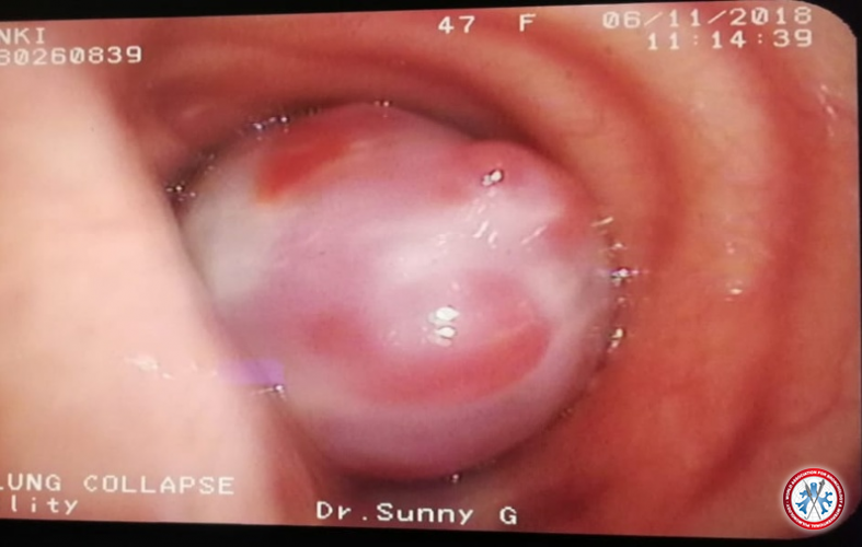
47 year old female presenting with features of Left lung collapse clinically and Chest X ray wise with a similar Chest X ray at hand showing the same findings.Bronchoscopic appearance of a pearly ehite glistening polypoidal intraluminal growth with smooth glistening white surface and very vascular lesion is characteristic of Bronchial Carcinoid.
Image courtesy of
DR.Sunny George MD,DNB,FRCP,FCCP
imagecontest Screenshot 2018 11 06 21 51 36 532 com.whatsapp
imagecontest Screenshot 2018 11 06 21 51 36 532 com.whatsapp
-
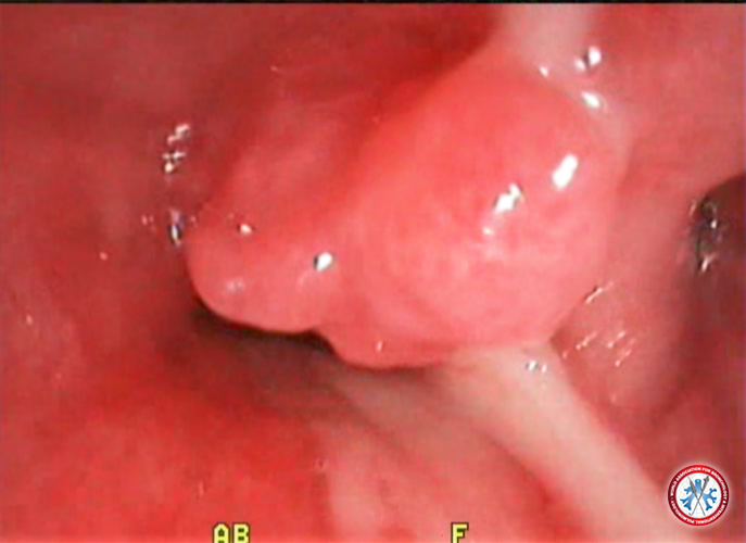
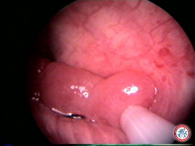
RT Bronchial Fibroepithelial Polyp (rare cae) Treatment was complete Resection with cryo
Image courtesy of Ayman Rizk, M.D.
5bcf5b8610650 RT Bronchial Polyp 2
5bcf5b8610650 RT Bronchial Polyp 2
-
Granulation tissue with partial obstruction of distal trachea after multiple EBUS TBNA attempts to a firm sarcoid 4R node
Image courtesy of Amir Abramovich, MD
granulation
granulation
-
Fibroepithelial polyp . Diastal bronchus intermedius
Image courtesy of Amir Abramovich, M.D.
fibriephitelial polyp
fibriephitelial polyp
-
Endobronchial myxoma . RUL bronchus
Image courtesy of Amir Abramovich, M.D.
myxoma
myxoma
-
Giant Simple carcinoid obstructing RMB in carinal level
Image courtesy of Amir Abramovich, MD
a100S0002
a100S0002
-
Melanoma that lost its pigmantation obrtructing RUL
Image courtesy of Amir Abramovich, MD
a100S0006
a100S0006
-
Chronic pulmonary cavitary Asperagilosis . plug obstructing RB6
Image courtesy of Amir Abramovich, MD
57bee6441fdc6 100S0009
57bee6441fdc6 100S0009
-
Endobronchial MELANOMA in subsegment of LINGULA
Image courtesy of Amir Abramovich, MD
57bc66797d972 melanoma
57bc66797d972 melanoma
-
RUL obstruction by Bronchocentric granulomatosis
Image courtesy of Amir Abramovich, MD
RUL obstruction
RUL obstruction
-
Squamous cell ca obstructing coming from RUB and obstructing RMB
Image courtesy of Amir Abramovich, MD
RUB and obstructing RMB
RUB and obstructing RMB
-
RML Partial obstruction by Hemartoma with puss from post obstruction pneumonia
Image courtesy of Amir Abramovich, MD
RML Partial obstruction
RML Partial obstruction
-
Simple carcinoid bleeding spontaneously obstructing RML
Image courtesy of Amir Abramovich, MD
obstructing RML
obstructing RML
-
Bronchus intermedius Obstruction by metastatic Renal cell CA
Image courtesy of Amir Abramovich, MD
Bronchus intermedius
Bronchus intermedius
-
Close up on Glandular papiloma
Image courtesy of Amir Abramovich, MD
RUB 2 2
RUB 2 2
-
Glandular Papiloma at RUB inlet
Image courtesy of Amir Abramovich, MD
RMB 2 1
RMB 2 1
-
Simple carcinoid with wide base coming from posterior wall of RUB and obstructing it
Image courtesy of Amir Abramovich, MD
57a4560749a82 100S0002 3 1
57a4560749a82 100S0002 3 1
-
POST OBSTRUCTION BRONCHIECTASIS
Image courtesy of Amir Abramovich, MD
57a44ed480206 SUBSEGMENTAL LBG BRONCHIECTASIS AND PUSS 1 3
57a44ed480206 SUBSEGMENTAL LBG BRONCHIECTASIS AND PUSS 1 3
-
Mucos gland adenoma obstructing lb6 with post obstruction bronchiectasis on CT
Image courtesy of Amir Abramovich, MD
57a44ed47d666 LLB OBSTRACTING LESION 1 1
57a44ed47d666 LLB OBSTRACTING LESION 1 1
-
Endotracheal chondroma
Image courtesy of Hervé Dutau, MD
IMAGE10
IMAGE10
-
Post-lung transplantation anastomotic stenosis
Image courtesy of Hervé Dutau, MD
IMAGE9
IMAGE9
-
Post-intubation tracheal tear
Image courtesy of Hervé Dutau, MD
IMAGE8
IMAGE8
-
Pseudotumoral tuberculosis
Image courtesy of Hervé Dutau, MD
IMAGE7
IMAGE7
-
Endotracheal polypoid angioma
Image courtesy of Hervé Dutau, MD
image6
image6
-
Tracheobronchopathia osteochondroplastica
Image courtesy of Hervé Dutau, MD
Image3
Image3
-
Endobronchial sarcoidosis
Image courtesy of Hervé Dutau, MD
IMAGE5
IMAGE5
-
Post-radiation necrosis of the right lateral lower part of the trachea
Image courtesy of Hervé Dutau, MD
IMAGE1
IMAGE1
-
Tracheal amyloidosis
Image courtesy of Hervé Dutau, MD
IMAGE2
IMAGE2
-
Relapsing polychondritis
Image courtesy of Hervé Dutau, MD
image 4
image 4
-
7 years old boy with Bronchial Carcinoid Tumor in his Right Main Bronchus
Image courtesy of Mohammad Ashkan Moslehi, MD
Bronchial Carcinoid Tumor in his Right Main Bronchus
Bronchial Carcinoid Tumor in his Right Main Bronchus
-
TRACHEAL STENOSIS POST TRACHEOTOMY
Image courtesy of J. Pablo Diaz-Jimenez, MD
56e6c879b0c8f tracheal senosis posttracheotomy
56e6c879b0c8f tracheal senosis posttracheotomy
-
BENIGN FISTULA AFTER RADIOTHERAPY
Image courtesy of J. Pablo Diaz-Jimenez, MD
56e6c879af7dc Benign Esoeph tracheal fistulae
56e6c879af7dc Benign Esoeph tracheal fistulae
-
TRACHEAL STENOSIS POSTTRACHEOSTOMY
Image courtesy of J. Pablo Diaz-Jimenez, MD
56e6c71ebac5f tracheal senosis posttracheotomyq
56e6c71ebac5f tracheal senosis posttracheotomyq
-
Metastasis Melanoma LMSB
Image courtesy of J. Pablo Diaz-Jimenez, MD
metastasis melanoma LMSB
metastasis melanoma LMSB
-
Hamartoma LMSB
Image courtesy of J. Pablo Diaz-Jimenez, MD
LMSB hamartoma
LMSB hamartoma
-
Pre-procedure: PITS - Far to close views
Image courtesy of Kedar Hibare, MD
56ce91e9a38cc 1
56ce91e9a38cc 1
-
Neurofibroma
Image courtesy of Fabien Maldonado, MD
Neurofibroma
Neurofibroma
-
Recurrent respiratory papillomatosis
Image courtesy of Fabien Maldonado, MD
RRP
RRP
-
Hamartoma
Image courtesy of Fabien Maldonado, MD
hamartoma
hamartoma
-
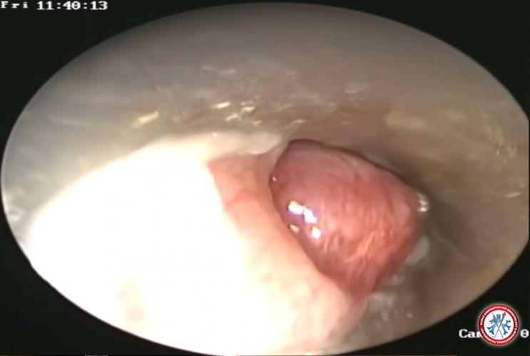
Carcinoid Tumor
Image courtesy of Fabien Maldonado, MD
Carcinoid
Carcinoid
-
Amyloidoma
Image courtesy of Fabien Maldonado, MD
Amyloidoma
Amyloidoma
-
Glomic tumor obstructing the upper trachea
Image courtesy of Hervé Dutau, MD
Glomic tumor obstructing the upper trachea
Glomic tumor obstructing the upper trachea
-
Pseudotumor obstructing the left main stem bronchus
Image courtesy of Hervé Dutau, MD
Pseudotumor obstructing the left main stem bronchus
Pseudotumor obstructing the left main stem bronchus
-
Obstructing the right lower lobe
Image courtesy of Hervé Dutau, MD
Obstructing the right lower lobe
Obstructing the right lower lobe
-
Leiomyoma obstructing the right lower lobe
Image courtesy of Hervé Dutau, MD
leiomyome2
leiomyome2
-
Adenoma obstructing the right lower lobe
Image courtesy of Hervé Dutau, MD
Adenoma obstructing the right lower lobe
Adenoma obstructing the right lower lobe
-
Lipoma on the posterior wall of the trachea
Image courtesy of Hervé Dutau, MD
Lipome
Lipome
-
Endobronchial metastasis of rectal cancer
Image courtesy of Hervé Dutau, MD
MTS rectum
MTS rectum
-
Endobronchial metastasis of adrenal gland sarcoma
Image courtesy of Hervé Dutau, MD
MTS corticosur nalome
MTS corticosur nalome
-
Malignant external compression secondary to thyroid cancer
Image courtesy of Hervé Dutau, MD
External compression of the trachea secondary to thyroid cancer
External compression of the trachea secondary to thyroid cancer
-
Complex Multi-level Subglottic and Upper Tracheal Fibrotic Stenosis
Image courtesy of Henri Colt, MD
Le Tracheal Stenosis Stenosis
Le Tracheal Stenosis Stenosis
-
Subglottic Stenosis from Active Wegener Granulomatosis
Image courtesy of Henri Colt, MD
wegener subglottis
wegener subglottis
-
Near Total Trachael Obstruction by Infliltrating and Nodular Thyroid Cancer
Image courtesy of Henri Colt, MD
thyroid
thyroid
-
Midtrachael Obstruction by polylobulated Mucoepidermoid Tumor
Image courtesy of Henri Colt, MD
mucoepidermoid
mucoepidermoid
-
Mid-tracheal stenosis from inflitrating adenoid cystic carcinoma
Image courtesy of Henri Colt, MD
adenoid cystic carcinoma
adenoid cystic carcinoma
-
Squamous cell carcinoma obstructing the left main stem bronchus
Image courtesy of Hervé Dutau, MD
SCC
SCC


